Read more
A single-volume resource for detailed coverage of the anatomy, function, and pathology of the cranial nerves with CT and MRI correlation
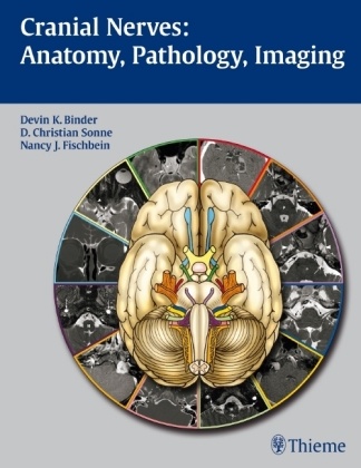
This beautifully illustrated book combines a detailed exposition of the anatomy and function of the cranial nerves with practical coverage of clinical concepts for the assessment and differential diagnosis of cranial nerve dysfunction. An introductory chapter provides a brief overview of cranial nerve anatomy and function, skull base anatomy, classification of pathologies, and imaging approaches. Each of the twelve chapters that follow is devoted to in-depth coverage of a different cranial nerve. These chapters open with detailed discussion of the various functions of each nerve and normal anatomy. The authors then describe common lesions and present a series of cases that are complemented by CT images and MRIs to illustrate disease entities that result in cranial nerve dysfunction.
Features
- Concise descriptions in a bulleted outline format enable rapid reading and review- Tables synthesize key information related to anatomy, function, pathology, and imaging- More than 300 high-quality illustrations and state-of-the-art CT and MR images demonstrate important anatomic concepts and pathologic findings- Pearls emphasize clinical information and key imaging findings for diagnosis and treatment- Appendices include detailed information on brainstem anatomy, pupil and eye movement control, parasympathetic ganglia, and cranial nerve reflexes
This book is an indispensable reference for practicing physicians and trainees in neurosurgery, neurology, neuroradiology, radiology, and otolaryngology-head and neck surgery. It will also serve as a valuable resource for students seeking to gain a solid understanding of the anatomy, function, and pathology of the cranial nerves.
List of contents
The 12 cranial nerves will each have their own individual chapter. Each chapter will cover anatomic information regarding the origin, course, and function of the cranial nerve, followed by differential diagnosis of the various pathologies affecting the nerve accompanied by imaging examples with associated description. Each chapter will include 1-3 line drawings, 3-12 radiographs of normal anatomy, and 8-30 radiographs of pathology.
About the author
Associate Professor of Radiology and, by courtesy, Otolaryngology-Head and Neck Surgery, Neurology, and Neurosurgery, Department of Radiology, Stanford University, Stanford, CA, USA
Summary
A single-volume resource for detailed coverage of the anatomy, function, and pathology of the cranial nerves with CT and MRI correlationThis beautifully illustrated book combines a detailed exposition of the anatomy and function of the cranial nerves with practical coverage of clinical concepts for the assessment and differential diagnosis of cranial nerve dysfunction. An introductory chapter provides a brief overview of cranial nerve anatomy and function, skull base anatomy, classification of pathologies, and imaging approaches. Each of the twelve chapters that follow is devoted to in-depth coverage of a different cranial nerve. These chapters open with detailed discussion of the various functions of each nerve and normal anatomy. The authors then describe common lesions and present a series of cases that are complemented by CT images and MRIs to illustrate disease entities that result in cranial nerve dysfunction.FeaturesConcise descriptions in a bulleted outline format enable rapid reading and review Tables synthesize key information related to anatomy, function, pathology, and imaging More than 300 high-quality illustrations and state-of-the-art CT and MR images demonstrate important anatomic concepts and pathologic findings Pearls emphasize clinical information and key imaging findings for diagnosis and treatment Appendices include detailed information on brainstem anatomy, pupil and eye movement control, parasympathetic ganglia, and cranial nerve reflexesThis book is an indispensable reference for practicing physicians and trainees in neurosurgery, neurology, neuroradiology, radiology, and otolaryngology-head and neck surgery. It will also serve as a valuable resource for students seeking to gain a solid understanding of the anatomy, function, and pathology of the cranial nerves.

