Read more
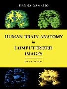
Zusatztext In Dr. Damasio's thoroughly revised second edition of her book! she enhances the understanding of common principles of gross brain organization. It provides users with confidence in their ability to identify focal regions of cerebral cortex on computerized scans! and leads them through the intricacies of the relationship of gross morphology to Brodmann architecture. This outstanding atlas thus facilitates further detailed exploration of the functional attributesof the cerebral cortical structures so carefully analyzed throughout the work. Informationen zum Autor Hanna Damasio is Distinguished Professor of Neurology at The University of Iowa College of Medicine! where she directs the Human Neuroanatomy and Neuroimaging Laboratory! and Adjunct Professor at the Salk Institute. Using computerized tomography and magnetic resonance scanning! she developed methods of investigating human brain structure and studied functions such as language using both the lesion method and functional neuroimaging. She is the author of over 150scientific publications and of the award-winning Lesion Analysis in Neuropsychology (Oxford University Press)! which has been used worldwide in brain-imaging work. Damasio is a Fellow of the American Academy of Arts and Sciences and of the American Neurological Association. She recently shared theSignoret Prize in cognitive neurosciences with Antonio Damasio. She holds honorary doctorates from the Universities of Lisbon and Aachen. Since 1992 she has been listed in Best Doctors in America under Neurology. Klappentext Spectacular recent developments in neuroimaging technologies have vastly increased the amount of information about brain structure that can be obtained from tomographic scans. Prepared by a leading expert in advanced brain-imaging techniques! this unique atlas illustrates the wide range of neuroanatomical variation in a collection of normal human brains in three-dimensional computerized reconstructions of MR scans of living persons. It also provides 100 sections of a single brain so that the same structure presented in the section of one incidence can be identified in the section of another incidence that intersects it. Axial and coronal sections of another brain with a different overall configuration are included at the two most frequently used incidences so that readers will get a sense of the "correction" that they may need to apply to standard images. The atlas is based on a voxel-rendering technique developed in the author's laboratory that permits the reconstruction of the brain in three dimensions with about the same degree of precision in identifying major sulci and gyri that can be achieved at the autopsy table. The images used throughout the atlas have not been beautified; the contours have been left ragged for greater anatomical detail. Thirteen pages of color illustrations are also included. The first of its kind! this atlas will be an essential tool for neurologists! neurosurgeons! neuroradiologists and neuroscientists! as well as for medical and neuroscience students. Zusammenfassung Neuroimaging technologies reveal the structure of the human brain in detail. This poses a problem: how to identify a particular brain site! be it normal or damaged by disease. This book shows the localization of brain structures that illustrate the range of neuroanatomical variation. It contains images from brain specimens. ...

