Share
Fr. 90.00
Rik Brydson, RMD Brydson, Rik Brydson, Brydson Rik
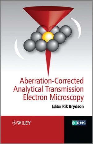
Aberration-Corrected Analytical Electron Microscopy
English · Hardback
Shipping usually within 3 to 5 weeks
Description
Informationen zum Autor Professor R.M.D. (Rik) Brydson is based in the School of Process, Environmental and Materials Engineering at the University of Leeds, UK. He is a committee member for the European Microscopy Society as well as the Electron Microscopy and Analysis Group (Institute of Physics). Klappentext Electron microscopy has undergone significant developments in recent years due to the practical implementation of schemes which can diagnose and correct for the imperfections (aberrations) in both the probe-forming and the image-forming electron lenses. This book presents the background and implementation of techniques which have allowed true imaging and chemical analysis at the scale of single atoms as applied to the fields of materials science and nanotechnology.Edited and written by the founders of the world's first aberration corrected Scanning Transmission Electron Microscope facility (SuperSTEM at Daresbury Laboratories in the UK), this text:* Presents the theory, instrumentation and applications of aberration correction in transmission electron microscopes* Is based on an established course taught at postgraduate summer schools by leaders in this field.* Is essential reading for researchers involved in the analysis of materials at the nanoscaleIdeal for final-year undergraduates and postgraduate students, as well as academics and industrialists involved in electron microscopy, this book can be used as a component of courses in nanotechnology, materials science, physics, chemistry or engineering disciplines. Zusammenfassung The book is concerned with the theory, background, and practical use of transmission electron microscopes with lens correctors that can correct the effects of spherical aberration. The book also covers a comparison with aberration correction in the TEM and applications of analytical aberration corrected STEM in materials science and biology. Inhaltsverzeichnis List of Contributors xi Preface xiii 1 General Introduction to Transmission Electron Microscopy (TEM) 1 Peter Goodhew 1.1 What TEM Offers 1 1.2 Electron Scattering 3 1.2.1 Elastic Scattering 7 1.2.2 Inelastic Scattering 8 1.3 Signals which could be Collected 10 1.4 Image Computing 12 1.4.1 Image Processing 12 1.4.2 Image Simulation 13 1.5 Requirements of a Specimen 14 1.6 STEM Versus CTEM 17 1.7 Two Dimensional and Three Dimensional Information 17 2 Introduction to Electron Optics 21 Gordon Tatlock 2.1 Revision of Microscopy with Visible Light and Electrons 21 2.2 Fresnel and Fraunhofer Diffraction 22 2.3 Image Resolution 23 2.4 Electron Lenses 25 2.4.1 Electron Trajectories 26 2.4.2 Aberrations 27 2.5 Electron Sources 30 2.6 Probe Forming Optics and Apertures 32 2.7 SEM, TEM and STEM 33 3 Development of STEM 39 L.M. Brown 3.1 Introduction: Structural and Analytical Information in Electron Microscopy 39 3.2 The Crewe Revolution: How STEM Solves the Information Problem 41 3.3 Electron Optical Simplicity of STEM 42 3.4 The Signal Freedom of STEM 45 3.4.1 Bright-Field Detector (Phase Contrast, Diffraction Contrast) 45 3.4.2 ADF, HAADF 45 3.4.3 Nanodiffraction 46 3.4.4 EELS 47 3.4.5 EDX 47 3.4.6 Other Techniques 48 3.5 Beam Damage and Beam Writing 48 3.6 Correction of Spherical Aberration 49 3.7 What does the Future Hold? 51 4 Lens Aberrations: Diagnosis and Correction 55 Andrew Bleloch and Quentin Ramasse 4.1 Introduction 55 4.2 Geometric Lens Aberrations and Their Classification 59 4.3 Spherical Aberration-Correctors 66 4.3.1 Quadrupole-Octupole Corrector 69 4.3.2 Hexapole Corrector 70 4.3.3 Parasitic...
List of contents
List of Contributors xi
Preface xiii
1 General Introduction to Transmission Electron Microscopy (TEM) 1
Peter Goodhew
1.1 What TEM Offers 1
1.2 Electron Scattering 3
1.2.1 Elastic Scattering 7
1.2.2 Inelastic Scattering 8
1.3 Signals which could be Collected 10
1.4 Image Computing 12
1.4.1 Image Processing 12
1.4.2 Image Simulation 13
1.5 Requirements of a Specimen 14
1.6 STEM Versus CTEM 17
1.7 Two Dimensional and Three Dimensional Information 17
2 Introduction to Electron Optics 21
Gordon Tatlock
2.1 Revision of Microscopy with Visible Light and Electrons 21
2.2 Fresnel and Fraunhofer Diffraction 22
2.3 Image Resolution 23
2.4 Electron Lenses 25
2.4.1 Electron Trajectories 26
2.4.2 Aberrations 27
2.5 Electron Sources 30
2.6 Probe Forming Optics and Apertures 32
2.7 SEM, TEM and STEM 33
3 Development of STEM 39
L.M. Brown
3.1 Introduction: Structural and Analytical Information in Electron Microscopy 39
3.2 The Crewe Revolution: How STEM Solves the Information Problem 41
3.3 Electron Optical Simplicity of STEM 42
3.4 The Signal Freedom of STEM 45
3.4.1 Bright-Field Detector (Phase Contrast, Diffraction Contrast) 45
3.4.2 ADF, HAADF 45
3.4.3 Nanodiffraction 46
3.4.4 EELS 47
3.4.5 EDX 47
3.4.6 Other Techniques 48
3.5 Beam Damage and Beam Writing 48
3.6 Correction of Spherical Aberration 49
3.7 What does the Future Hold? 51
4 Lens Aberrations: Diagnosis and Correction 55
Andrew Bleloch and Quentin Ramasse
4.1 Introduction 55
4.2 Geometric Lens Aberrations and Their Classification 59
4.3 Spherical Aberration-Correctors 66
4.3.1 Quadrupole-Octupole Corrector 69
4.3.2 Hexapole Corrector 70
4.3.3 Parasitic Aberrations 72
4.4 Getting Around Chromatic Aberrations 74
4.5 Diagnosing Lens Aberrations 75
4.5.1 Image-based Methods 77
4.5.2 Ronchigram-based Methods 80
4.5.3 Precision Needed 85
4.6 Fifth Order Aberration-Correction 85
4.7 Conclusions 86
5 Theory and Simulations of STEM Imaging 89
Peter D. Nellist
5.1 Introduction 89
5.2 Z-Contrast Imaging of Single Atoms 90
5.3 STEM Imaging Of Crystalline Materials 92
5.3.1 Bright-field Imaging and Phase Contrast 93
5.3.2 Annular Dark-field Imaging 96
5.4 Incoherent Imaging with Dynamical Scattering 101
5.5 Thermal Diffuse Scattering 103
5.5.1 Approximations for Phonon Scattering 104
5.6 Methods of Simulation for ADF Imaging 106
5.6.1 Absorptive Potentials 106
5.6.2 Frozen Phonon Approach 107
5.7 Conclusions 108
6 Details of STEM 111
Alan Craven
6.1 Signal to Noise Ratio and Some of its Implications 112
6.2 The Relationships Between Probe Size, Probe Current and Probe Angle 113
6.2.1 The Geometric Model Revisited 113
6.2.2 The Minimum Probe Size, the Optimum Angle and the Probe Current 115
6.2.3 The Probe Current 115
6.2.4 A Simple Approximation to Wave Optical Probe Size 117
6.2.5 The Effect of Chromatic Aberration 117
6.2.6 Choosing ±opt in Practice 118
6.2.7 The Effect of Making a Small Error in the Choice of ±opt 119
6.2.8 The Effect of ± On the Diffraction Pattern 120
6.2.9 Probe Spreading and Dep
Product details
| Authors | Rik Brydson, RMD Brydson |
| Assisted by | Rik Brydson (Editor), Brydson Rik (Editor) |
| Publisher | Wiley, John and Sons Ltd |
| Languages | English |
| Product format | Hardback |
| Released | 23.09.2011 |
| EAN | 9780470518519 |
| ISBN | 978-0-470-51851-9 |
| No. of pages | 296 |
| Series |
RMS - Royal Microscopical Society RMS - Royal Microscopical Soci RMS - Royal Microscopical Society RMS - Royal Microscopical Soci |
| Subjects |
Natural sciences, medicine, IT, technology
> Chemistry
Chemie, Physik, Mikroskopie, Elektronenmikroskopie, Microscopy, chemistry, Physics, Optik u. Photonik, Optics & Photonics |
Customer reviews
No reviews have been written for this item yet. Write the first review and be helpful to other users when they decide on a purchase.
Write a review
Thumbs up or thumbs down? Write your own review.

