Share
Fr. 111.00
Klaus Dieter Budras, Klaus-Diete Budras, Klaus-Dieter Budras, Paul R. Greenough, Robert Habel, Robert E Habel...
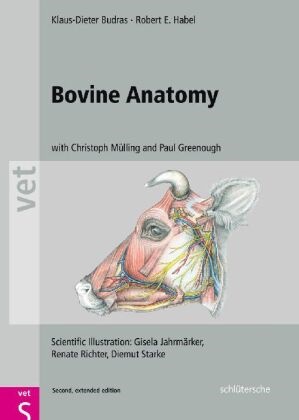
Bovine Anatomy
English · Hardback
Shipping usually within 1 to 3 working days
Description
Die zweite englische Auflage dieses erfolgreichen Lehrbuches ist nun auch nach dem bewährten Konzept der "Budras-Atlanten" durch namhafte Experten aus der Anatomie und der klinischen Medizin um die klinisch-funktionelle Anatomie erweitert."This is a much-needed textbook-atlas that depicts bovine anatomy. It is appropriately organized such that it can easily be the single book that veterinarians refer to when an anatomic question needs to be answered about this species. It is most definitely worth the price."JAVMA - Journal of the American Veterinary Medical Association
List of contents
Preface
Topographic Anatomy
Thoracic limb (A. Wünsche, R. Habel and K.-D. Budras)
Skeleton of the thoracic limb
Muscles and nerves of the shoulder, arm, and forearm
Cutaneous nerves, blood vessels, and lymph nodes of the thoracic limb
Vessels and nerves of the manus
Interdigital nerves and vessels, interossei, and fasciae of the manus
Synovial structures of the thoracic limb
Pelvic limb (A. Wünsche, R. Habel and K.-D. Budras)
Skeleton of the pelvic limb
Lateral thigh and cranial crural muscles with their nerves
Medial thigh and caudal crural muscles with their nerves
Cutaneous nerves, blood vessels, and lymph nodes of the pelvic limb
Arteries, veins, and nerves of the pes
Dermis of the hoof (Ch. Mülling and K.-D. Budras)
The hoof (ungula) (Ch. Mülling and K.-D. Budras)
Synovial structures of the pelvic limb (Ch. Mülling and K.-D. Budras)
Head (R. Habel, and K.-D. Budras)
Skull and hyoid apparatus (R. Habel and K.-D. Budras)
Skull with teeth (R. Habel and K.-D. Budras)
Skull with paranasal sinuses and horns (R. Habel and K.-D. Budras)
Superficial veins of the head, facial n. (VII), and facial muscles (S. Buda and K.-D. Budras)
Trigeminal n. (V3 and V2), masticatory mm., salivary gll., and lymphatic system (S. Buda and K.-D. Budras)
Accessory organs of the eye (P. Simoens and K.-D. Budras)
The eyeball (bulbus oculi) (P. Simoens and K.-D. Budras)
Nose and nasal cavities, oral cavity and tongue (S. Buda, R. Habel, and K.-D. Budras)
Pharynx and larynx (S. Buda, R. Habel and K.-D. Budras)
Arteries of the head and head-neck junction, the cran. nn. of the vagus group (IX–XI), and the hypoglossal n. (XII)(S. Buda and K.-D. Budras)
Central nervous system and cranial nerves
The brain (R. Habel and K.-D. Budras
Cranial nerves I–V (S. Buda, H. Bragulla and K.-D. Budras)
Cranial nerves VI–XII (S. Buda, H. Bragulla, and K.-D. Budras)
Spinal cord and autonomic nervous system (S. Buda and K.-D. Budras)
Vertebral column, thoracic skeleton, and neck (A. Wünsche, R. Habel and K.-D. Budras)
Vertebral column, ligamentum nuchae, ribs, and sternum
Neck and cutaneous muscles
Deep shoulder girdle muscles, viscera and conducting structures of the neck
Thoracic cavity
Respiratory muscles and thoracic cavity with lungs (Ch. Mülling and K.-D. Budras)
Heart, blood vessels, and nerves of the thoracic cavity (R. Habel and K.-D. Budras)
Abdominal wall and abdominal cavity
The abdominal wall (R. Habel, A. Wünsche and K.-D. Budras)
Topography and projection of the abdominal organs on the body wall
Stomach with rumen, reticulum, omasum, and abomasum (A. Wünsche and K.-D. Budras)
Blood supply and innervation of the stomach; lymph nodes and omenta (R. Habel, A. Wünsche and K.-D. Budras)
Spleen, liver, pancreas, and lymph nodes (P. Simoens, R. Habel and K.-D. Budras)
Intestines with blood vessels and lymph nodes (P. Simoens, R. Habel and K.-D. Budras)
Pelvic cavity and inguinal region, including urinary and genital organs
Pelvic girdle with the sacrosciatic lig. and superficial structures in the pubic and inguinal regions (R. Habel and K.-D. Budras)
Inguinal region with inguinal canal, inguinal lig., and prepubic tendon (R. Habel and K.-D. Budras)
Lymphatic system, adrenal glands, and urinary organs (K.-D. Budras and A. Wünsche)
Arteries, veins, and nerves of the pelvic cavity (A. Wünsche and K.-D. Budras)
Female genital organs (H. G. Liebich and K.-D. Budras)
The udder (H. Bragulla, H. König, and K.-D. Budras)
The udder with blood vessels, lymphatic system, nerves, and development (H. Bragulla, H. König, and K.-D. Budras)
Male genital organs and scrotum (R. Habel and K.-D. Budras).
Perineum, pelvic diaphragm, ischiorectal fossa, and tail (R. Habel and K.-D. Budras)
Anatomical aspects of bovine spongiform encephalopathy (BSE) (S. Buda, K.-D. Budras, T. Eggers, R. Fries, R. Habel, G. Hildebrandt, K. Rauscher, and P. Simoens)
Special Anatomy, Tabular Part
Myology
Lymphatic system
Peripheral nervous system
Contributions to Clinical-Functional Anatomy
Applied anatomy of the carcass (K.-D. Budras, R. Fries, and R. Berg)
References
Index
About the author
Professor Klaus-Dieter Budras ist Lehrstuhlinhaber des Instituts für Veterinär-Anatomie in Berlin.
Summary
Die zweite englische Auflage dieses erfolgreichen Lehrbuches ist nun auch nach dem bewährten Konzept der „Budras-Atlanten“ durch namhafte Experten aus der Anatomie und der klinischen Medizin um die klinisch-funktionelle Anatomie erweitert.
„This is a much-needed textbook-atlas that depicts bovine anatomy. It is appropriately organized such that it can easily be the single book that veterinarians refer to when an anatomic question needs to be answered about this species. It is most definitely worth the price.”
JAVMA – Journal of the American Veterinary Medical Association
Additional text
…a ‘must-have’ book for anyone with an interest in bovine medicine… not only provides a detailed atlas of bovine anatomy; it also includes a comprehensive section on clinical anatomy… consistent layout of pages, with text on the left and drawings on the right… The large size is beneficial, the drawings are very clear and the illustration of structures from different angles makes a good visual aid for dissection; the colours are eye-catching and make it a pleasure just to leaf through the book… The coverage of material is comprehensive while remaining clinically relevant… The quality of the images and illustrations is superb and the descriptions of procedures are extremely helpful… very reasonable price
–Renate Weller, Kate Holroyd, Andrea Turner and Peter Aitken, Veterinary Record, 22-Oct-2011
Report
a must-have book for anyone with an interest in bovine medicine not only provides a detailed atlas of bovine anatomy; it also includes a comprehensive section on clinical anatomy consistent layout of pages, with text on the left and drawings on the right The large size is beneficial, the drawings are very clear and the illustration of structures from different angles makes a good visual aid for dissection; the colours are eye-catching and make it a pleasure just to leaf through the book The coverage of material is comprehensive while remaining clinically relevant The quality of the images and illustrations is superb and the descriptions of procedures are extremely helpful very reasonable price Renate Weller, Kate Holroyd, Andrea Turner and Peter Aitken Veterinary Record 20111022
Product details
| Authors | Klaus Dieter Budras, Klaus-Diete Budras, Klaus-Dieter Budras, Paul R. Greenough, Robert Habel, Robert E Habel, Robert E. Habel, Gisela Jahrmärker, Chri Mülling, Christoph K. W. Mülling, Renate Richter, Diemut Starke |
| Assisted by | Gisela Jahrmärker (Illustration), Renate Richter (Illustration), Diemut Starke (Illustration) |
| Publisher | Schlütersche |
| Languages | English |
| Product format | Hardback |
| Released | 01.12.2022 |
| EAN | 9783899930528 |
| ISBN | 978-3-89993-052-8 |
| No. of pages | 176 |
| Dimensions | 252 mm x 18 mm x 350 mm |
| Weight | 1333 g |
| Illustrations | Illustr. |
| Series |
Schlütersche Vet |
| Subjects |
Natural sciences, medicine, IT, technology
> Medicine
> Veterinary medicine
Skelett, Anatomie, Rind, Kopf, Körper, Hals, TOPOGRAPHISCHE ANATOMIE, ZNS, schultergelenk, Körperregionen, Schultergliedmaße, BECKENHÖHLE |
Customer reviews
No reviews have been written for this item yet. Write the first review and be helpful to other users when they decide on a purchase.
Write a review
Thumbs up or thumbs down? Write your own review.

